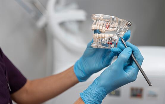
Cone-beam computed tomography 3D is the most informative method for diagnosing the dental system, facial skeleton, as well as the paranasal, maxillary and paranasal sinuses among x-ray studies.
3D computed tomography of two jaws (upper and lower) with recording on disk
40 BYN
3D computed tomography of one jaw
3D computed tomography of one jaw (upper or lower) with recording on disk
25 BYN
3D computed tomography of the TMJ
3D computed tomography of the TMJ with closed/open mouth with disk recording
50 BYN
Advantages of 3D tomography
- High accuracy: CBCT provides a complete and detailed image, which helps doctors provide the most effective treatment.
- Safety: Radiation exposure is significantly lower than with classic CT. The procedure is safe for adults and children.
- Fast: The scanning itself takes only 20–40 seconds, and the results are available immediately.
- Convenience: Modern equipment ensures a comfortable and painless examination.
- Full digital documentation: Images can be saved electronically, sent to your doctor, or used for treatment planning.
How is the CBCT procedure performed?
The procedure for performing dental x-rays is simple and safe for the patient. Using cone beam tube technology, the X-ray beam passes exclusively through a small portion of the area being examined, scanning it. The data is then processed by special software and converted into an incredibly high-resolution 3D image.
The 3D tomography obtained in this way can be studied literally in different planes: rotated and viewed from any angle, made virtual “slices” to study the smallest details, zoomed in, and made the necessary measurements.
The 3D dental image data is recorded on a disk, which is provided to the patient, and in the future any dentist has the opportunity to view and study it on a regular computer in his clinic.
The area of study can be the entire maxillofacial region, for which it is necessary to take a panoramic photograph of the teeth in Vitebsk, or individual segments that require study. In this case, a photograph of the area of interest in the dental system is taken.
Extent of radiation exposure during cone beam tomography
The amount of radiation exposure during the procedure depends on the type of equipment and scanning parameters. Due to the small size of the treatment area, the patient receives a minimal dose of radiation. Radiation exposure during CBCT is only 40-60 μSv, which makes it possible to safely conduct such studies several times a year! Safety for the patient is also ensured by compliance with safety precautions. When used correctly and following medical protocols, CBCT of the jaw is a completely safe and effective diagnostic tool. To take a 3D photo of your teeth in Vitebsk, you must first contact your dentist for a referral.
CBCT result
The result of a 3D image of the teeth is recorded on a disk and given to the patient. Your doctor will be able to open the disk on his computer.
Areas of application of CBCT
- Otorhinolaryngology - diagnosis of odontogenic, non-odontogenic inflammatory diseases and neoplasms of the maxillary sinuses
- Endodontics - identifying periodic changes and their location, counting the number of root canals and roots, quality control of root canal filling
- Periodontology - assessment of the degree of bone resorption of the alveolar process
- Orthodontics - construction of cephalometric sections in lateral and frontal projections, for children, diagnosis of changes in baby teeth and anomalies in the development of the dental system
- Gnathology - viewing in a 3D volumetric image and in three planes of the temporomandibular joints, assessing the width of the joint space and the structure of the bone tissue of the articular head
- Implantology - implantation planning by accurately measuring the height and width of the alveolar ridge in the planned area, choosing an implant based on the data obtained and its intended location
- Orthopedics - planning and prognosis of orthopedic treatment
- Surgery - study of the location of pathological changes in the alveolar processes of the jaws, maxillary sinuses and mandibular canals; the location of impacted teeth and their relationship with surrounding structures. In case of injuries to teeth and jaws, assessment of the location, nature of the fracture and displacement of fragments.
Book an appointment
Request sent
Our specialist will contact you shortly!
Сообщение не отправлено
К сожалению, что-то сломалось при отправке сообщения
Comprehensive diagnostics and treatment in ophthalmology, dentistry, ENT diseases, functional diagnostics (ECG, ultrasound), as well as 3D tomography (CBCT), laboratory tests, etc.
Call: +375 29 640 77 00
Book an appointment
Request sent
Our specialist will contact you shortly!
Сообщение не отправлено
К сожалению, что-то сломалось при отправке сообщения